Research
Re-characterisation of ‘non-specific’ anterior knee pain after total knee replacements by the diagnosis of patella mal-tracking using a novel ultrasound method in the clinic
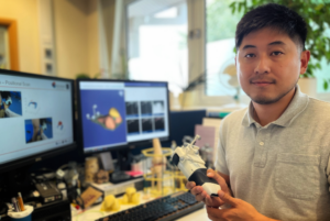
Jack Tu is a post-doctoral research fellow in Clinical Biomechanics at the Oxford Orthopaedic Engineering Centre within The Nuffield Department of Orthopaedics, Rheumatology and Musculoskeletal Sciences and recipient of Orthopaedic Research UK’s Early Career Research Fellowship. He is using a novel joint dynamic analysis approach to understand non-specific anterior knee pain after total knee replacement (TKR).
‘Some people continue to experience pain after a knee replacement, and we need better methods to confirm the cause,’ Tu described. After total knee replacement, up to one in five patients report persistent non-specific anterior knee pain, which can impact their quality of life and mental health. Abnormal patella tracking, where the kneecap moves out of place when the leg straightens or bends, is suspected to be the cause of pain. However, what constitutes ‘normal’ patella tracking is not yet defined, nor are there reliable tools to quantify patella tracking.
TKR usually focuses on the femur and tibia. Despite the kneecap also being important, it is often ignored or not the focus during treatment. ‘If we don’t understand the problem, we won’t be able to do anything efficiently,’ said Tu. For patients opting to have revision surgery, the outcome may not be as good if there is not a clear understanding of the problem. ‘That costs a lot of money to the medical system,’ the researcher explained.
The initial aims of the research are to directly observe and measure patella tracking, to define the distribution of ‘normal’ tracking among TKR patients and to define thresholds for abnormal tracking associated with pain, dissatisfaction, and poor outcomes. The long-term goal is to optimise patient diagnosis and post-operative care. Tu hopes the system will help clinicians identify patients who might benefit from early physiotherapy interventions and reduce the proportion of patients undergoing revision surgery that may not improve their outcome.
Tu works within an interdisciplinary team led by Associate Prof Stephen Mellon and Prof David Murray, whose primary focus is combining approaches to develop techniques to improve joint replacement. ORUK has previously supported a project to develop the approach – Dynamic Musculoskeletal Ultrasound Measurement of Joint Pain. The new project will further develop the technology and explore how its use in the clinic can benefit patients.
Tu described his system: ‘I’m using ultrasound combined with motion analysis to provide a new kind of joint dynamic analysis which cannot be provided by traditional scans such as X-ray and MRI.’
Clinical motion capture is used in rehabilitation to assess performance or as a pre-surgical assessment for children with cerebral palsy, but it is not currently used to assess problems with knee joints. ‘No one has done this because there are some limitations for joint dynamics using motion analysis,’ Tu added. The researcher described, ‘If you try to touch your kneecap and bend then straighten your knee, you will feel the kneecap is actually moving around underneath your skin. This almost makes tracking the kneecap with traditional motion analysis impossible, as you have to attach several markers, usually reflective balls, to the skin.’ He continued, ‘The combined use of motion capture and ultrasound allows us to observe and measure the motion of the kneecap.’
The system will not be tied to one manufacturer or device. Tu said, ‘I think every hospital has access to ultrasound systems, and some have a motion capture system. Although our system currently works with wireless portable probes from one manufacturer, we have developed the software to be able to receive images from any ultrasound system and data from any motion capture system so that we can do assessments anywhere. Our goal is to make the system economical to build and accessible to all.’ The research fellow mentioned, ‘We are also looking at some different possibilities, for example, a more mobile device, which is more attractive for some business partners. For now, we are using the VICON motion capture system because it’s reliable and extensively used, so we can access it in our local hospital. But we have trialled the system with other motion capture systems, and we are looking at some other options from the robotics world to make the system even more portable. In the future, I think as long as you have the right device, you will be able to use our system.’
This project builds on the previous lab-based project. ‘I’m spending some time before this project to prepare the system and to show that this is something we can actually bring into a clinical room,’ Tu shared. He explained, ‘What I’m trying to do in this ethically approved project is to recruit some people with and without pain after their surgery. We are using the new method combined with ultrasound and the motion analysis system to look at their dynamic movement – the relationship between the kneecap and thigh.’ Participants are asked to do some typical daily activities, and researchers will attempt to correlate specific kneecap movement patterns to the incidence of pain.
‘The idea of combining ultrasound and motion capture data for the purposes of research has been there for some time, but until now, processing assessment data took many hours. In the past, the procedure was so long: after the data collection, researchers were still spending lots of time working on processing the image, combining it with the motion analysis and then generating analytical data. Now, I’ve been able to combine everything in one place; it’s semi-automatic, so it saves lots of human labour,’ Tu revealed.
The studies aim to test whether the system can be applied in the real world. The researcher expanded, ‘So, it’s not just me working with the data and creating some simulations but real clinically relevant results from patients. The performance of the system in the clinic and the opportunity to develop the system to work seamlessly in that environment is invaluable in terms of where we want to go with it in the future.’
Additionally, Tu hopes the system can have applications beyond knee replacement. ‘We can make it available for other clinical groups, and it is not limited to joint replacement because knee problems are also an area of interest for physios, sports clinics, or during growth in the younger generation. With our system, everyone should have the ability to quickly pick up an ultrasound scanner and do a dynamic assessment of problem joints. We are also interested in branching out, for example, to have applications at the shoulder and the hip joints in the future.’
The idea of combining ultrasound and motion capture data for the purposes of research has been there for some time, but until now, processing assessment data took many hours…Now, I’ve been able to combine everything in one place; it’s semi-automatic, so it saves lots of human labour.

Tell us your thoughts